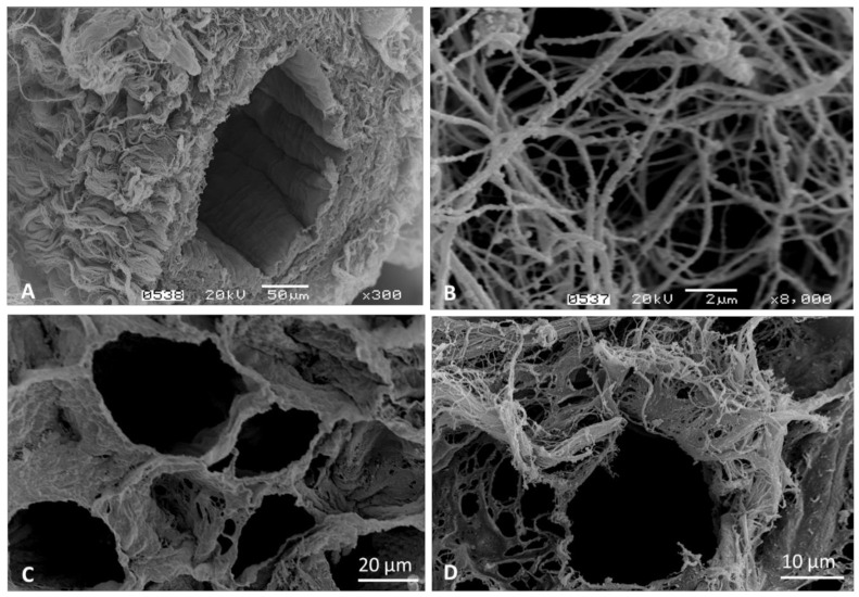
Figure 3.
Scanning electron microscopy imaging of the bovine decellularized bone marrow. (A) Intact vascular structure, (B) Reticular fibers in connective tissue, (C) Adipose tissue ECM, preserved cellular niches, (D) Individual cell niche. The images are part of the Hematology and Hemotherapy Center archive of the characterization of the decellularized bone marrow (DeBM) [18]; however, these images have not been published elsewhere.

