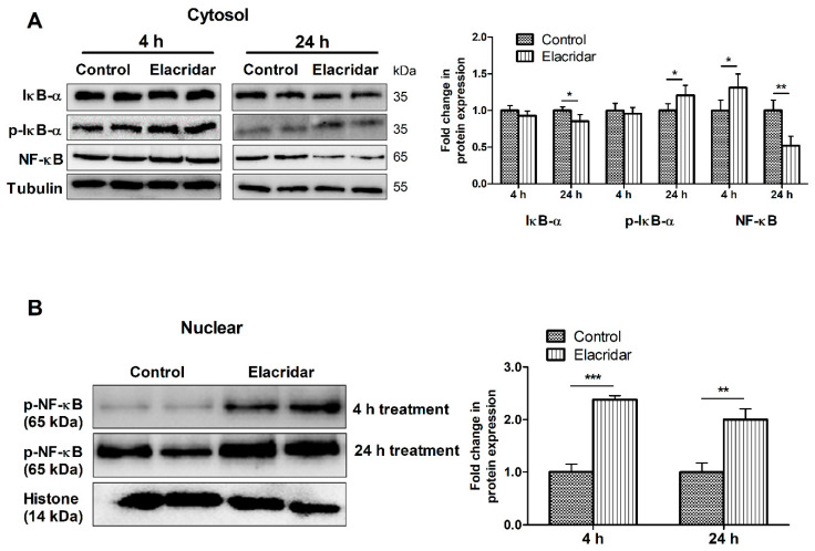
Figure 2.
Treatment with elacridar (5 µM) significantly activated the NF-κB pathway in cultured bEnd3 cells in vitro. (A) Representative Western blot and densitometry analysis of the cytosolic fraction of IκB-α, p-IκB-α, and NF-κB, presented as fold change by elacridar compared to vehicle treatment, at 4 and 24 h post-treatment. (B) Representative Western blot and densitometry analysis of the nuclear fraction of p-NF-κB, presented as fold change by elacridar compared to vehicle treatment, at 4 and 24 h post-treatment. Statistical analysis was determined by Student’s t-test. Data are presented as mean ± SD of 3 independent experiments. * p < 0.05, ** p < 0.01, *** p < 0.001 compared to control. kDa indicates the molecular weight of analyzed proteins.

