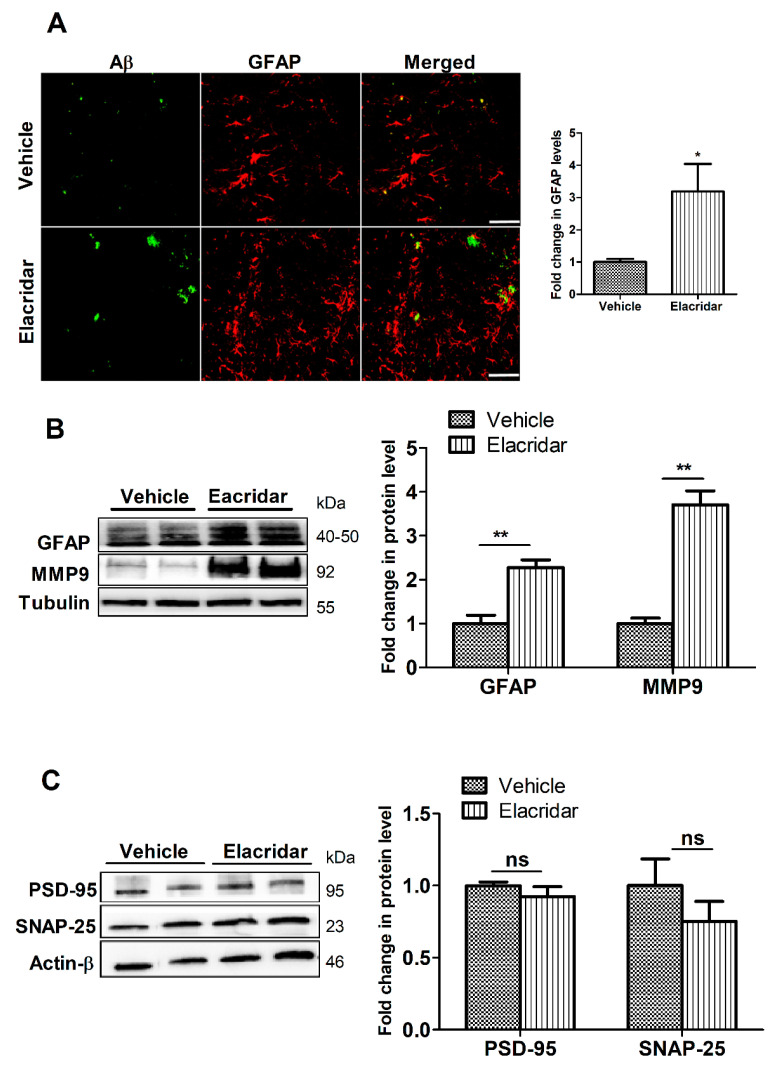
Figure 6.
Elacridar treatment (10 mg/kg/day i.p. for 28 days) significantly increased astrogliosis marker GFAP in TgSwDI mouse brains. (A) Representative brain sections from mouse hippocampus stained with GFAP antibody (red) to stain activated astrocytes and with 6E10 (green) antibody to detect total Aβ, with the corresponding quantification of GFAP. Scale bar, 50 μm. The semi-quantification analysis is presented as fold change caused by elacridar when compared to vehicle treatment. (B) Representative Western blot and densitometry analysis of GFAP expressions in mouse brain homogenates. (C) Representative Western blot and densitometry analysis of PSD-95 and SNAP-25 expressions in mouse brain homogenates. Data from Western blot is presented as fold change by elacridar on each protein compared to vehicle treatment. Statistical analysis was determined by Student’s t-test. Data are presented as mean ± SEM for n = 5 mice per group. ns = not significant, * p < 0.05, ** p < 0.01, compared to vehicle-treated group. kDa indicates the molecular weight of analyzed proteins.

