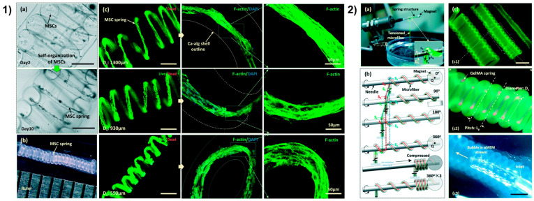
Figure 6.
3D vSM from [196] with permission. (1) Self-organization of cells in GelMA spring (a), 13.33 length-to-diameter ratio of a MSC spring (b), live/dead images of MSCs spring in GelMA spring (c), F-actin (green) and nuclei (blue) staining of MSCs spring. Unlabeled scale bars 400 µm. (2) Semi-automated coiling assembly (a,b), images of helical microtubes (c) with various diameters (c1), GelMA spring (red) in the helical microtube of Ca-alginate shell (green) (c2), perfusion of the helical microtube (c3). Scale bars 800 µm.

