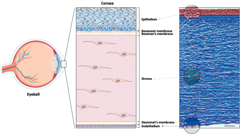
Figure 1.
Schematic of the human cornea and histology. Left panel: Schematic view of the human eye. The cornea forms the transparent front part of the eyeball. Central panel: From anterior to posterior, the cornea is made up of a stratified squamous epithelium deposited on a basement membrane, follows the Bowman’s membrane, a stroma, composed predominantly of collagen fibrils in which keratocytes are entangled, the Descemet’s membrane, and a monolayer of endothelial cells. Right panel: Masson trichrome staining of a section of the entire native human cornea showing all cellular compartments of that tissue. Scale bar: 50 μm.

