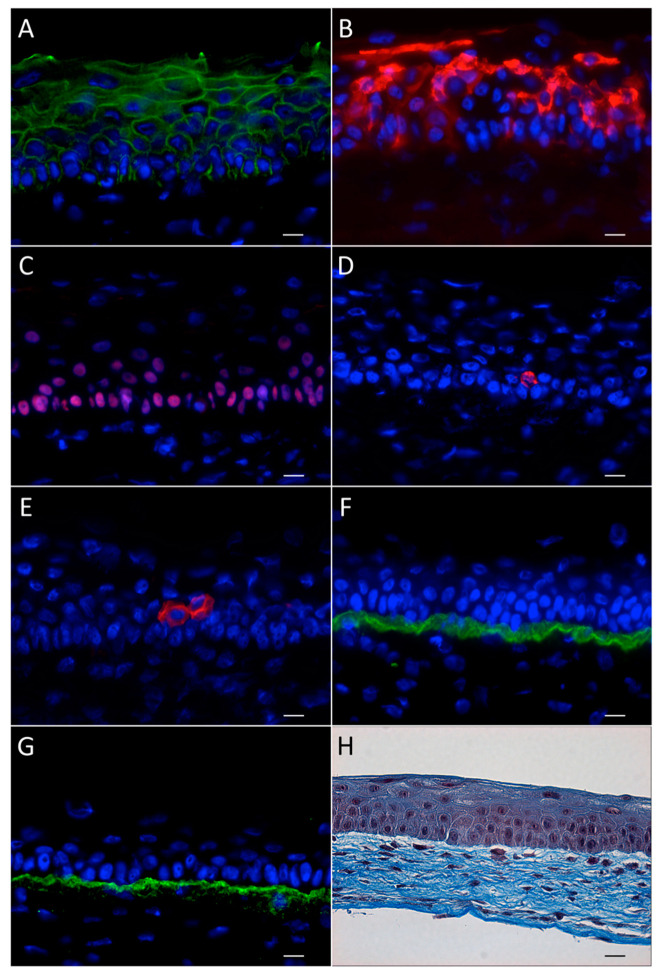
Figure 3.
Characterization of the human tissue-engineered cornea. Immunofluorescence analysis of: (A) the epithelial barrier marker ZO-1, (B) the differentiation marker keratins K3/K12, (C) the epithelial cell marker p63, (D) the cornea epithelial stem cell marker ΔNp63α, and (E) K19, (F) the epithelial basement membrane components laminin V, and (G) collagen IV. (H) Nuclei were counterstained with Hoechst 33,258 reagent and appear in blue. Scale bar: 20 µm. Histology (Masson’s Trichrome staining) of the tissue-engineered cornea, showing an epithelium adhered to the self-assembled stromal matrix. The 5–6 epithelial cell layers differentiate during their upward migration. The scale bar in H equals 20 µm.

