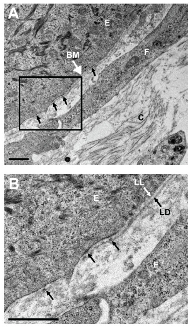
Figure 4.
Transmission electron microscopic analysis of the hTEC. (A) Electron microscopic examination of the hTEC revealed the presence of an organized basement membrane (BM) with many hemidesmosomes (arrows) that attach basal corneal epithelial cells (E) to the underlying fibroblast sheet (F). A basement membrane is present at the junction between the epithelium and the stroma. Note the presence of the collagen fibers (C) surrounding the fibroblasts in the sheet. (B) Higher magnification that shows both the lamina lucida (LL) and lamina densa (LD), as well as the hemidesmosomes (arrows) are present in the BM. Scale bars: 1 µm.

