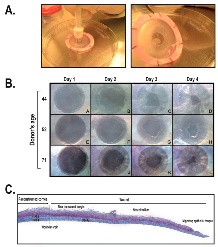
Figure 5.
Production of wounds on human tissue-engineered corneas. (A) The reconstructed cornea is wounded using an 8 mm biopsy punch. (B) Closure of wounds on tissue-engineered corneas produced using HCECs isolated from the eyes of three different donors (44, 52, and 71 years old). Closure of the wounded epithelia was followed from 1 to 4 days after corneal injury. (C) Composite image showing a complete view of the wounded tissue-engineered human cornea 3 days following corneal damage (sections were stained with Masson’s trichrome; cells are pink and collagen is bluish). The wound margin created by the biopsy punch is indicated. F1 + F2: initial fibroblast sheets present in the reconstructed cornea prior to wounding. F3 + F4: supplementary fibroblast sheets added following wounding of the tissue-engineered corneas (Figure adapted from Reference [173] with the permission of the journal Biomaterials).

