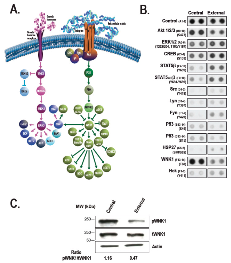
Figure 7.
Kinase profiling analysis during corneal wound healing. (A) Major protein mediators from the WNK1 signal transduction pathways. (B) Cell lysates isolated from the central and external areas of hTECs assembled using hCEC-44, hCEC-52, and hCEC-71 were pooled together and used for the detection of activated kinases with the Human Phospho-Kinase Array from R&D Systems. Kinases and mediators identified as being differentially phosphorylated between the central (wounded) and external (unwounded) areas of hTECs are identified. (C) Cell lysates from the central and external areas of wounded hTECs were analyzed by immunoblotting to confirm the phosphokinase array results for the mediator WNK1. Actin was used as the loading control (Figure adapted from Reference [336] with the permission of Journal of Tissue Engineering and Regenerative Medicine).

