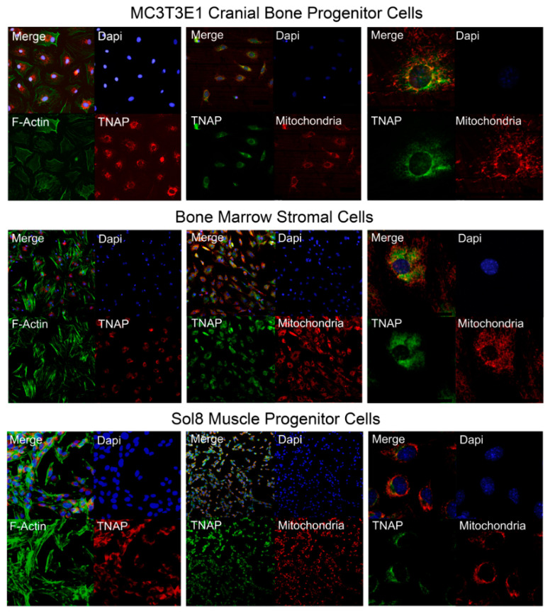
Figure 9.
TNAP is expressed in a peri-nuclear pattern and partially co-localizes with mitochondria in undifferentiated bone and muscle progenitor cells. Immunofluorescent staining of undifferentiated MC3T3E1 cranial osteoprogenitor cells, bone marrow stromal cells, and Sol8 muscle progenitor cells reveals that TNAP is localized internally at a peri-nuclear region, and is co-localized with mitochondria. Left panels: representative images of co-localization of TNAP (red) and F-Actin (green). Middle panels: representative images of co-localization of TNAP (green) and mitochondria (red). Right panels: representative images of co-localization of TNAP (green) and mitochondria (red) at a higher magnification. Nuclear stain was performed with DAPI (blue).

