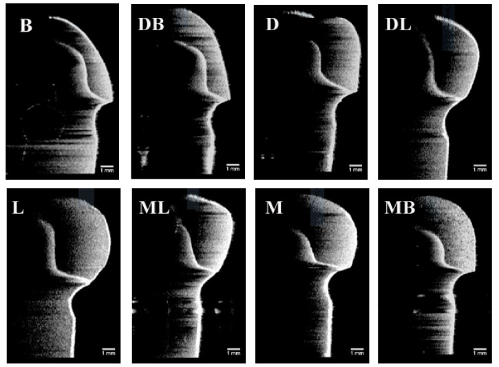
Figure 6.
Images of the optical coherence tomographic scans of a representative CAD/CAM provisional crown (Group CAM) at eight selected locations. (B) At buccal point, (DB) at distal-buccal point, (D) at distal point, (DL) at distal-lingual point, (L) at lingual point, (ML) at mesial-lingual point, (M) at mesial point, and (MB) at mesial-buccal point. All the orientations of the OCT images have been rotated by 90 degrees from the original orientation of the OCT images.

