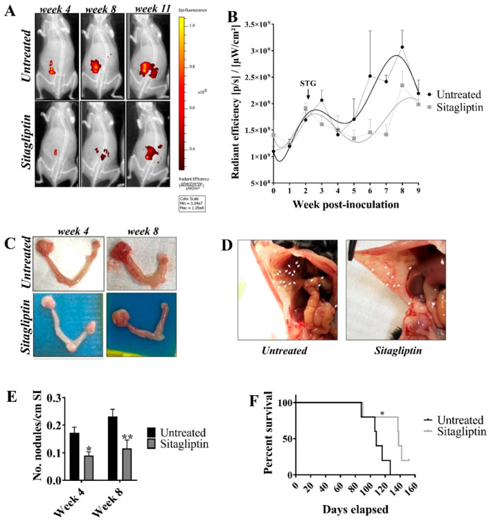
Figure 2.
Tumour burden and metastatic spread in ID8 pROSA-iRFP720 tumour-bearing mice following sitagliptin treatment. (A) ID8 pROSA-iRFP720 cells were implanted into the ovarian bursa and iRFP720 fluorescence was measured using the IVIS Lumina III In Vivo Imaging System (Perkin Elmer, Waltham, MA, USA). Mice were imaged at FOV C (n = 5–15/group) and iRFP720 fluorescence was detected using the iRFP filter set at an exposure time of 5 s. iRFP signal was isolated using a custom spectral unmixing algorithm. Representative images of iRFP fluorescence are shown. (B) Quantitative region of interest (ROI) analysis of iRFP total radiant efficiency [p/s]/[µW/cm2] over time. (C) Representative images of ovaries from untreated mice or mice receiving sitagliptin at weeks 4 and 8 post tumour inoculation. ID8 pROSA-iRFP720 tumours are shown on the right and nontumour bearing ovaries are shown on the left. (D) The proportion of macroscopic tumour nodules on the small intestine/length (cm). (E) Representative images of tumour nodules on the peritoneal wall from untreated mice and mice receiving sitagliptin treatment. (F) The Kaplan-Meier curve and log-rank test of overall survival analysis for ID8 pROSA-iRFP720 tumour-bearing mice. Data are presented as mean ± SD (upper SD for iRFP720 fluorescence). * = p < 0.05; ** = p < 0.01.

