Figure 5.
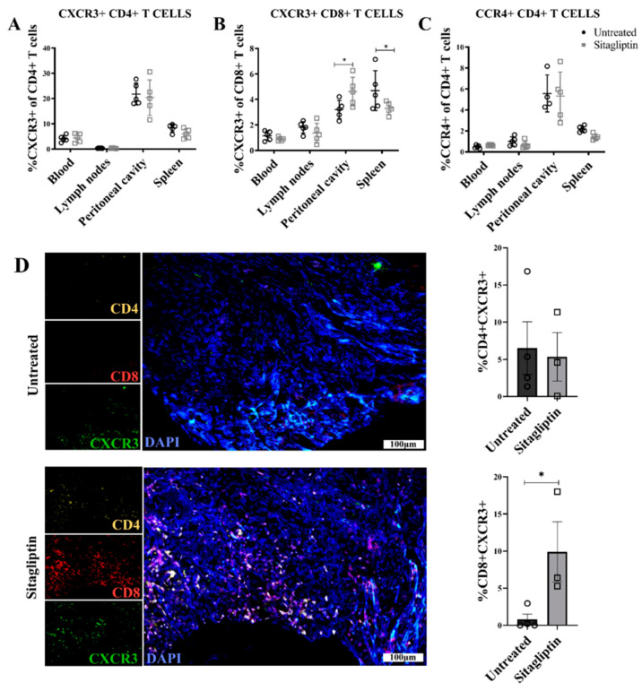
Chemokine receptor expression in ID8 tumour-bearing mice treated with sitagliptin. Leukocytes were isolated from the blood, lymph nodes, peritoneal cavity and spleen of ID8 pROSA-iRFP720-bearing mice at week four post-inoculation and examined using a BD LSRFortessa X-20 (BD Biosciences). Percentage of (A) CXCR3+ CD4+ T cells, (B) CD8+ T cells, and (C) CCR4+ CD4+ T cells of the corresponding parent population. (D) Representative images of ovarian tumour sections stained with CD4 (yellow), CD8 (red) and CXCR3 (green) at four weeks post tumour inoculation. Nuclei were stained with DAPI (blue). Bar graphs show the percentage area of ovarian tumour sections stained with (i) CD4+CXCR3+ and (ii) CD8+CXCR3+. Images were acquired using the VS120 Virtual Slide Microscope (Olympus Corporation, Japan) and processed using the Olyvia software v2.9.1 (Olympus Corporation, Japan). Data were analysed by calculating the percentage area of CD4+CXCR3 or CD8+CXCR3+ colocalisation of total tissue area using a consistent binary threshold in ImageJ v1.0 (National Institute of Health, MD, USA). * = p < 0.05.

