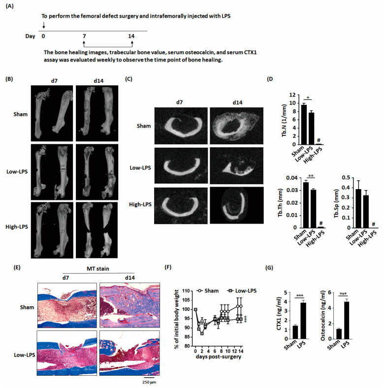
Figure 1.
Lipopolysaccharides (LPS) delayed new bone formation and bone healing in mice with femoral bone defects. (A) Schematic representation of the experimental setup. (B) Micro-computed tomography (microCT) three-dimensional image of the bone defect site show that low-dose LPS delays bone healing and high-dose LPS promotes bone fracture in the bone defect area. (C) Representative transverse images of the fractured femur by microCT. (D) LPS decreases the trabecular number and thickness. (E) MT stained histological sections demonstrate LPS inhibits bone bridge formation during bone healing. (F) LPS impairs body weight recovery. (G) LPS increases the serum levels of CTX1 and osteocalcin, an indicator of the bone turnover rate. Data are presented as the mean ± standard error of the mean. Analyses were conducted with two-way analysis of variance followed by Bonferroni’s post-hoc test. * p < 0.05, ** p < 0.01, *** p < 0.001. Abbreviations: LPS, lipopolysaccharide; Tb.N, trabecular number; Tb.Th, trabecular thickness; Tb.Sp, trabecular separation; MT, Masson’s trichrome stain; CTX1, C-terminal telopeptides of type I collagen; #, full fracture. Sham, n = 5; Low-LPS, n = 7; and High-LPS, n = 5.

