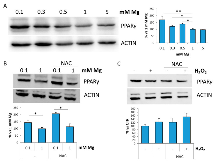
Figure 4.
Low magnesium induces PPARγ. (A) HUVEC were cultured in low (0.1, 0.3, 0.5 mM), physiological (1 mM) and high (5 mM) extracellular Mg for 24 h. (B) HUVEC were cultured in 0.1 or 1 mM in the presence or in the absence of NAC (5 mM) for 24 h. (C) HUVEC were treated with H2O2 (200 μM) for 30 min in the presence or in the absence of NAC (5 mM) and then maintained in culture for 24 h. Cell extracts were processed for Western blot using antibodies against PPARγ. Actin was used as a control of loading. A representative blot is shown. Densitometry (right panel) was performed on three different blots by Image J. * p ≤ 0.05 and ** p ≤ 0.01.

