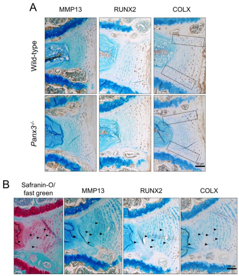
Figure 4.
Localization of hypertrophic chondrocyte markers in Panx3-/- IVDs. (A) Representative mid-sagittal sections of lumbar IVDs from 19-month-old WT and Panx3-/- mice immunostained for either MMP13, RUNX2 or COLX (indicated by brown stain). Sections were counterstained with methyl green. Black boxes highlight the CEP-AF interface (n = 5 mice per group, 4–6 IVDs per mouse). (B) Serial sections of a representative WT lumbar IVD at 19 months-of-age stained with either safranin-O/fast green (red stain indicative of proteoglycan content) or anti- MMP13, RUNX2 or COLX antibody (indicated by brown stain) counterstained with methyl green. Arrowheads indicate enlarged AF cells.

