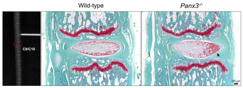
Figure 6.
Degenerative changes identified in adjacent uninjured NP tissue of Panx3-/- mice following needle puncture. Representative X-ray image demonstrating caudal IVD 8/9 undergoing needle puncture injury. Dotted red circle highlights caudal IVD 9/10 of the motion segment distal to the site of injury. Representative safranin-O/fast green-stained mid-sagittal sections of uninjured caudal IVDs distal to the punctured IVDs, harvested 6-weeks after injury. Images are representative of n = 6 mice per group. Black arrow indicates accelerated NP degeneration marked by increased matrix density and reduced cellularity.

