Figure 1.
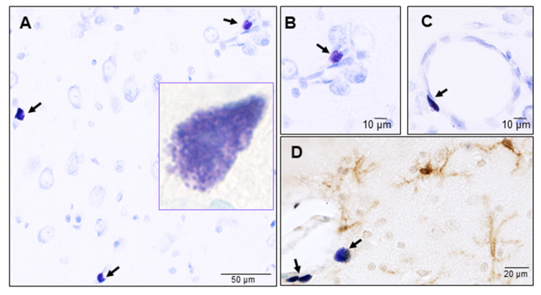
Mast cells in the naïve rat brain. Mast cells were identified by toluidine blue staining and their typical metachromasia. (A) Mast cells at the level of thalamus, inset shows a high magnification view of a granulated parenchymal mast cell; (B) A roundish mast cell around a small blood capillary; (C) An elongated mast cell in close proximity to a larger blood capillary; (D) Mast cells in close association with microglia (identified by an antibody against the ionized calcium binding adaptor molecule-1 (Iba-1) brown stain) in the brain parenchyma. Arrows indicate toluidine blue stained mast cells. Shown in panel D is a roundish mast cell and two elongated mast cells in close proximity to a blood capillary.

