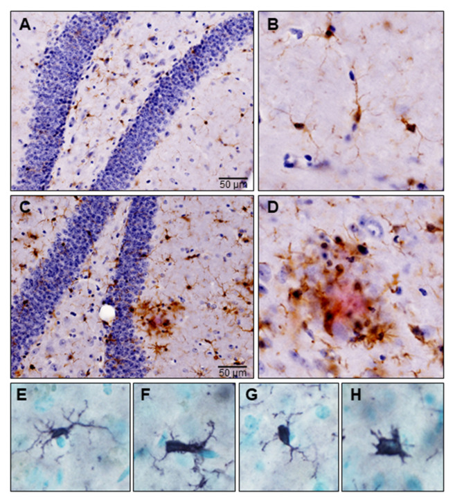
Figure 2.
Microglia in a triple transgenic mouse model of Alzheimer’s disease (3xTg-AD). Formalin-fixed free-floating sections were subjected to immunostaining using antibodies against Iba-1 and β-amyloid (clone 6E10). Microglia were identified by brown signal and amyloid plaques by red signal. (A) Microglia in the healthy mouse brain at the level of hippocampus; (B) High magnification image of ramified microglia with a small cell body and fine extensive processes; (C) Activated microglia are present throughout the hippocampal section; (D) High magnification image of activated microglia surrounding amyloid plaque; (E–H) Morphological transformation of homeostatic microglia to disease-associated phenotypes, where their branches appear short, stubby and cells appear amoeboid.

