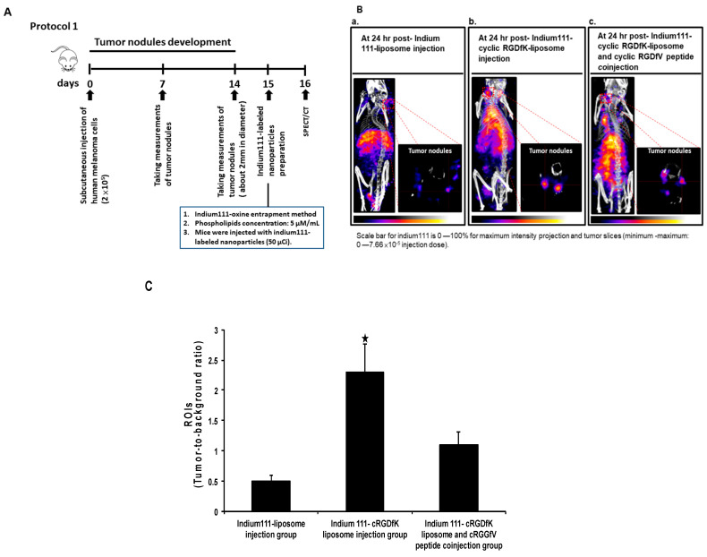
Figure 2.
Non-invasive imaging with Nano SPECT/CT in mice without a metastatic growth of human melanoma cells. (A) Experimental scheme; (B) The nude mice with human melanoma were injected with the 111In-liposome (panel a), with the 111In-labeled-cyclic RGDfK-liposome (panel b), or with an injection of being a combination of the 111In-labeled-cyclic RGDfK-liposome and the cyclic RGDfV peptide (panel c), and images was captured at 24 h post-radioactive liposomes injection. (C) The region of interest analysis of tumors was showed. The black star indicated p < 0.01, when compared to the other groups. The figure showed here was a representative example of three independent experiments in which each group consisted of three mice.

