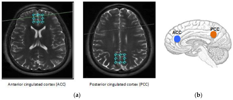
Figure 1.
Locations of the volume studied in the (a) anterior and (b) posterior cingulated cortices. The single voxel acquisition used a spin-echo sequence recorded within the following parameters: Echo time (TE) = 23 ms, repetition time (TR) = 1070 ms, 2 NEX, flip angle = 90°, and 256 acquisitions with the point-resolved spectroscopy (PRESS) technique. During data acquisition, the same experienced neuroradiologist, blind to the clinical data, placed the voxels (20 × 20 × 20 mm3) at the ACC and PCC.

