Figure 3.
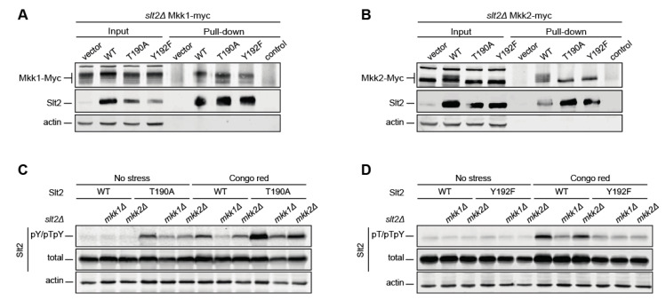
Analysis of Mkk1 and Mkk2 binding to differentially phosphorylatable forms of Slt2 and their effect on Slt2 phosphorylation. The strains slt2Δ MKK1-myc (YMJ30) (A) and slt2Δ MKK2-myc (YMJ31) (B) were transformed with the plasmid pRS316 alone (vector) or expressing, as indicated, Slt2 (WT), Slt2T190A, or Slt2Y192F. Cells were grown to the mid-exponential phase in YPD and then stimulated with 30 µg/mL of Congo Red (CR) for 2 h. Cell extracts (input) were incubated with the Mpk1 antibody attached to Dynabeads™ Protein G to purify Slt2 complexes (pull-down) and resolved by SDS–PAGE. Proteins were detected with anti-c-Myc, anti-Mpk1 (Slt2), and anti-actin antibodies by immunoblotting. The control of a Dynabeads-Mpk1 antibody non-incubated with cell extracts is included. (C,D) slt2Δ (Y00993 strain), mkk1Δ slt2Δ (YGGR32 strain), and mkk2Δ slt2Δ (YGGR33 strain) cells containing the same plasmids as in (A,B) were grown to the mid-exponential phase in YPD and then stimulated with 30 µg/mL of CR for 2 h. Cell extracts were resolved by SDS–PAGE, and analyzed by immunoblotting with anti-Slt2-pY/pTpY, anti-Slt2-pT/pTpY (as in Figure 1), anti-Mpk1 (total), and anti-actin antibodies. A representative assay from three different experiments with distinct transformants is shown.

