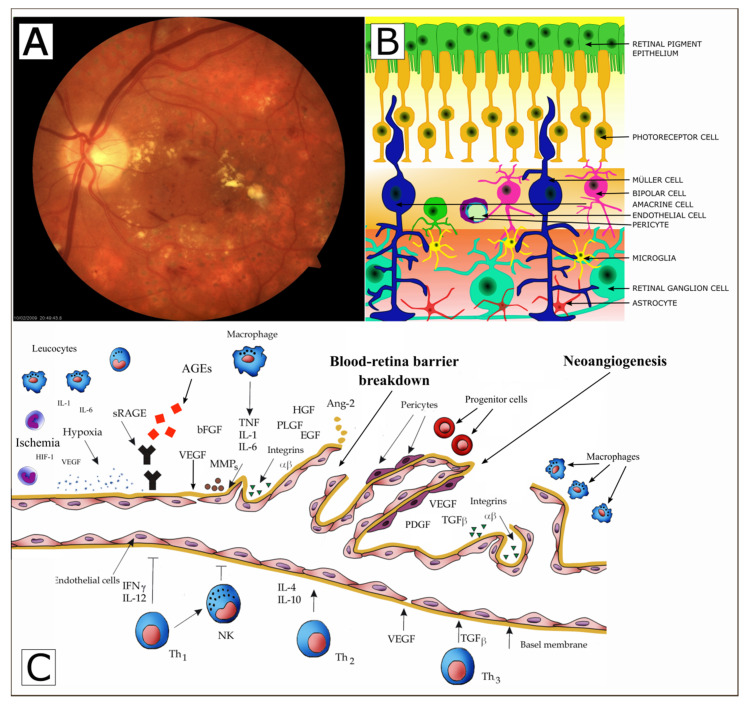
Figure 1.
Possible mechanism of retinal vascular endothelial cell dysfunction and neuroretinal degeneration in diabetic patients. Fundus photos of eyes from diabetic retinopathy accompanied by macular oedema (Panel A), modified from [42]. The neurovascular unit of the retina (retinal ganglion cells, Müller cells, microglia, astrocytes, endothelial cells, pericytes, and other). (Panel B), modified from [43]. The selected factors involved in the development and progression of diabetic retinopathy. AGEs—advanced glycation end products, RAGEs—receptor for advanced glycation end products, VEGF- vascular endothelial growth factor, IGF-I—insulin like growth factor, PLGF—placental growth factor, HGF—hepatocyte growth factor, PEDF pigment epithelium derived factor, bFGF—basic fibroblast growth factor, TGF-beta. transforming growth factor beta, MMPs—metalloproteinases PDGF-platelet-derived growth factor, EGF-Epidermal growth factor, Ang-2—Angiopoietin-2, (Panel C), modified from [42,43].

