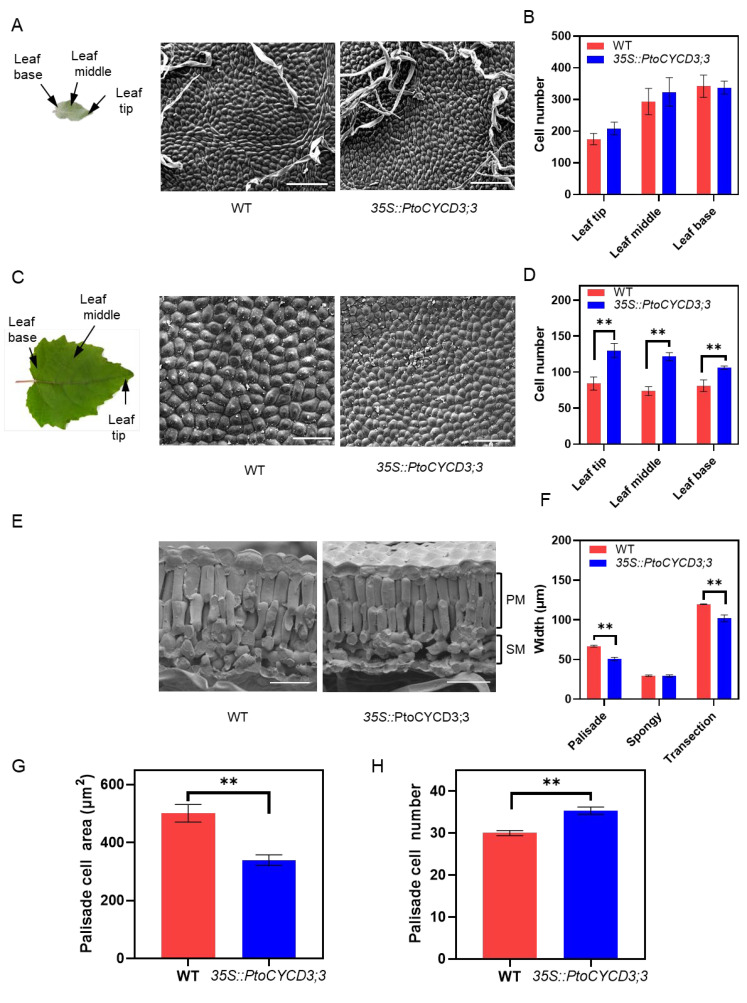
Figure 3.
Overexpression of PtoCYCD3;3 affect leaf development. (A) Schematic of a young leaf. Scanning electron micrographs of the adaxial epidermis of wild-type (WT) and 35S::PtoCYCD3;3 young leaves. (B) Number of cells per unit leaf area at the tip, middle, and base of WT and 35S::PtoCYCD3;3 young leaves. (C) Schematic of a mature leaf. Scanning electron micrographs of the adaxial epidermis of WT and 35S::PtoCYCD3;3 mature leaves. (D) Number of cells per unit leaf area at the tip, middle, and base of WT and 35S::PtoCYCD3;3 mature leaves. (E) Cryo-scanning electron micrographs of the cross-sections of WT and 35S::PtoCYCD3;3 leaves. PM: palisade mesophyll; SM: spongy mesophyll. (F) Palisade mesophyll, spongy mesophyll, and transection width of WT and 35S::PtoCYCD3;3 leaves. (G) PM cell area of WT and 35S::PtoCYCD3;3 leaves. (H) PM cell number per unit leaf area of WT and 35S::PtoCYCD3;3 leaves. Significance as determined by independent sample T-test: * p < 0.05, ** p < 0.01. The values are means ±SE, n = 3. Three transgenic lines were used. Bar, 100 µm (A,C); 40 µm (E).

