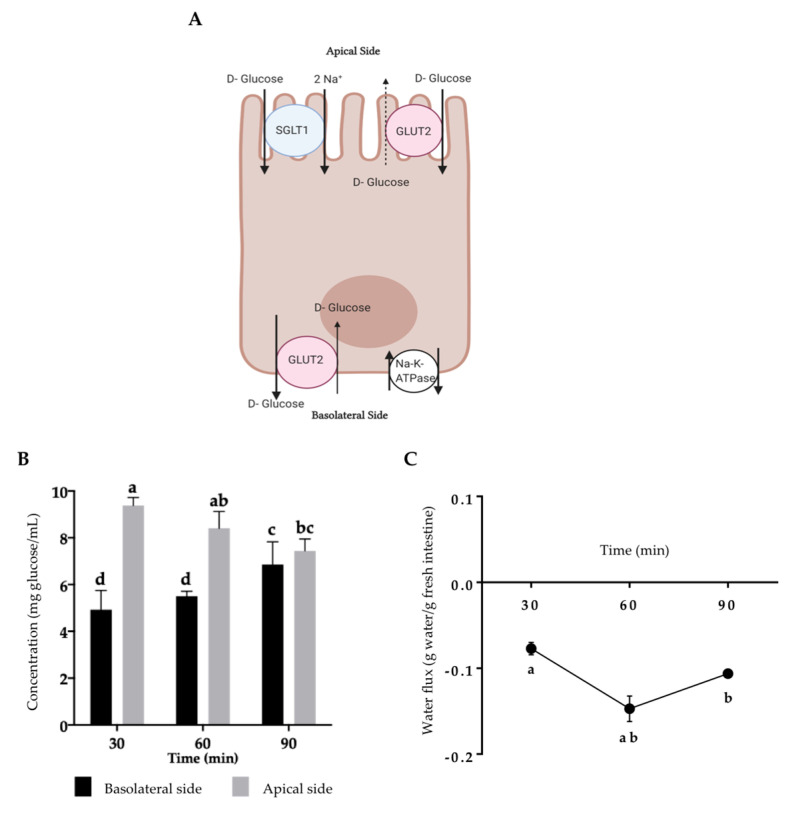
Figure 1.
Sprague–Dawley rats’ everted jejunum integrity during small intestine absorption. (A) Glucose transportation in enterocytes. (B) For the glucose flux determination, everted jejunum was incubated in glucose-free Krebs–Ringer buffer for 30, 60, and 90 min, and glucose was determined at 540 nm. (C) For the water flux determination, everted jejunum was incubated in Krebs–Ringer buffer for 30, 60, and 90 min, water was determined as a change in the gut sac weight. Results are expressed as the mean ± SD of three independent experiments. Small letters express significant differences between all the samples (p ≤ 0.05) by Tukey–Kramer’s test. The enterocyte model was created with BioRender® (https://app.biorender.com) and adapted from Koepsell [28].

