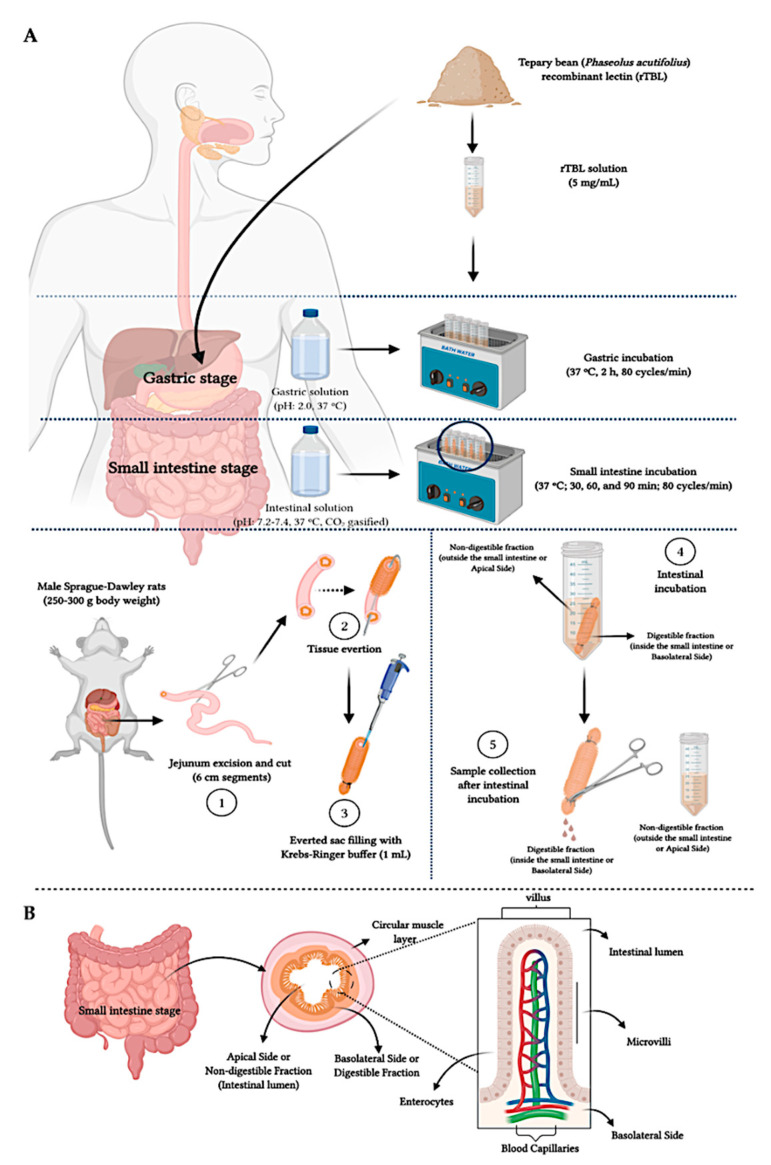
Figure 10.
Overall diagram of the in vitro/ex vivo gastrointestinal digestion. (A) Everted jejunum procedure. (1) The intestinal incubation was simulated ex vivo using a jejunum excised from male Sprague–Dawley rats, (2) carefully everted, (3) filled in the inner side (basolateral side) with Krebs–Ringer buffer (1 mL). Once tied on both sides, the filled intestinal tissue was placed in the intestinal solution (4) (pH adjusted sample to 7.2–7.4, added with intestinal enzymes: pancreatin and bile bovine) and incubated (30–60 min). Once incubated (5), the sample from the outer side of the small intestinal tissue was referred to as the non-digestible fraction (apical side), and the inner side of the intestinal tissue was considered as the digestible fraction (basolateral side). (B) Jejunum morphology.

