Figure 1.
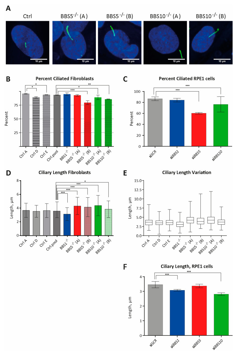
Investigation of the primary cilium in fibroblast obtained from patients with bardet biedl syndrome (BBS) and in the hTERT-immortalized retinal pigment epithelial cell-line, (RPE-1) transfected with small interfering RNA (siRNA) against the BBS genes. (A) IFM analysis of primary cilia. Primary cilia were labeled with anti-ARL13B antibody (green). Nuclei were visualized with DAPI staining (blue). Controls (Ctrl, here CtrlD) are also shown. Scale bars 10 µM. (B) Percentage of patient fibroblasts with cilia. Compared to percentage of ciliated cells in a pool of control fibroblasts (Ctrl.pool = CtrL.A + Ctrl.D + Ctrl.E; 92.60%, n = 798 cells), the percentage of ciliated cells was significantly lower in BBS5−/−(B) (79.54%, n = 308 cells, *** p =5.13 × 10⁻¹⁰), BBS10−/−(A) (89.16%, n = 369 cells, * p = 0.0494) and BBS10−/−(B) (85.78%, n = 225 cells, ** p = 0.0015) fibroblasts. No significant differences were observed for BBS1−/− (94.98%, n = 239, p = 0.202) and BBS5−/−(A) (92.77%, n = 249, p = 0.9301) compared to the pool of control fibroblasts. There was a significant difference in percentage of ciliated cells between the three controls (Ctrl). The percentage was significantly lower in Ctrl.D (89.18%, n = 305 cells) compared with Ctrl.A (95.72%, n = 234 cells; Ctrl.D/Ctrl.A: * p = 0.0054) and Ctrl.E (93.82%, n = 259 cells; Ctrl.D/Ctrl.E: * p = 0.037). No significant difference was observed between CtrlA and CtrlE (CtrlA/CtrlE: p = 0.34). (C) Percentage of siBBS RNA-treated RPE1 cells with cilia. The cilia were labeled with anti ARL13B antibody and nuclei were visualized with DAPI staining. For siRNA efficacy, see Supplementary Figure S1. Compared to percentage of ciliated cells in siSCR transfected RPE1 cells (86.81%, n = 379 cells), the percentage of ciliated cells were significantly decreased in RPE1 cells transfected with siBBS5 (60.20%, n = 294 cells, *** p = 2.2802 × 10⁻¹⁵) and siBBS10 (76.40%, n = 322 cells, *** p = 0.000347). No significant differences compared to siSCR transfected cells were obtained for siBBS1 (84.49%, n = 361 cells, p = 0.36789). (D) Quantification of primary cilia length in patient fibroblasts. No significant difference was obtained by comparing the three control fibroblasts lines (Ctrl.A, n = 224 cells; Ctrl.D, n = 270 cells; Ctrl.E n = 243 cells. Ctrl.A/Ctrl.D: p = 0.0918; Ctrl.A/Ctrl.E: p = 0.675; Ctrl.D/Ctrl.E: p = 0.207). A pool of all the controls (n = 737 cells) was used for comparison with the five BBS patient fibroblasts lines. Compared to the controls, BBS1−/− (n = 222, p = 3.77 × 10⁻¹¹) had significantly shorter cilia, whereas BBS5−/−(A) (n = 228, *** p = 1.83 × 10⁻¹³), BBS5−/−(B) (n = 242, *** p = 8.77 × 10⁻⁸), BBS10−/−(A) (n = 333, *** p = 2.00×10⁻¹⁶) and BBS10−/−(B) (n = 202, * p = 0.0165) all had significantly longer cilia. (E) Cilia length variation visualized as a boxplot of (C) showing the length variation for all cell-lines. Compared to control cell-lines, BBS5−/−(A), BBS5−/−(B) and BBS10−/−(A) had broader length distributions. (F) Quantification of primary cilia length in siBBS-treated RPE1 cells. The cilia length was significant shorter in RPE1 cells treated with siBBS1 (n = 361, *** p = 2.29 × 10⁻⁸), and siBBS10 (n = 322, *** p = 3.54 × 10⁻¹⁹) compared to siSCR-treated control RPE cells, whereas no significant difference was observed between siBBS5- (n = 294, p = 0.21) and siSCR-treated cells (n = 329). SiBBS transfection led to a substantial reduction in expression of the BBS genes (Supplementary Figure S1). In (B,C), the number of cells (n) investigated consist of pooled data from three separate experiments and significance was determined using the χ2-test. In (D,F), the number of cilia (n) measured consist of pooled data from three separate experiments. p-values and significance were determined using Student’s t-test, two-tailed. Significance levels p < 0.05, * p < 0.05, and *** p < 0.0005 were used. Error bars represent the standard deviation.

