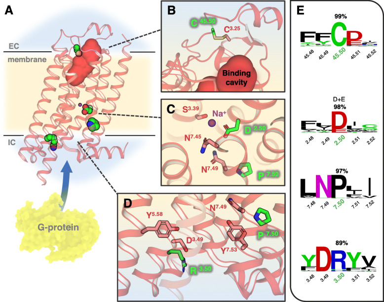
Fig. 8.
Structural visualization and examples of paradigmatic mutations selected from the study. a General view of the adenosine A2A receptor (PDBid: 4EIY) as a prototype GPCR with the TM boundaries indicated in light yellow and ECLs/ICLs regions in blue. The ligand-binding cavity is indicated by a solid red surface and G protein-interacting site by a blue arrow. Selected topological positions C45.50, D2.50, R3.50, and P7.50 are highlighted in the structure as green vdW spheres. b–d Closer look of the atomic environment of selected positions (green sticks) and surrounding residues (salmon). e Sequence conservation logos around the selected positions (number corresponds to their conservation percentage in the MSA). All the residue positions are referenced following the BW convention

