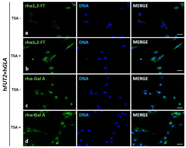
Figure 2.
Immunofluorescence analysis of in vitro-cultured porcine ACFCs treated (TSA+) (b,d) and not treated with trichostatin A (TSA−) (a,c). Representative microphotographs of immunofluorescence localization of recombinant human α1,2-fucosyltransferase (rhα1,2-FT; a,b) and α-galactosidase A (rhα-Gal A; c,d) in ACFCs derived from double-transgenic pigs (hFUT2×hGLA). Immunofluorescent staining with Alexa Fluor 488-labelled secondary antibodies (green fluorescence) and 4′,6-diamidino-2-phenylindole (DAPI)-mediated counterstaining of cell nuclei (blue fluorescence). Scale bars represent 100 μm. Immunoreaction was performed on in vitro cultured porcine ACFCs from at least three pigs of each experimental group. The immunofluorescence signal stemming from rhα1,2-FT was distributed in the perinuclear region of all the analyzed cells from each variant. The recombinant human α-galactosidase A was located homogeneously in whole cytoplasm of all the analyzed cells. The TSA+ cells exhibited a more intense signal for both rhα1,2-FT and rhα-Gal A proteins (b,d) than their TSA− counterparts (a,c).

