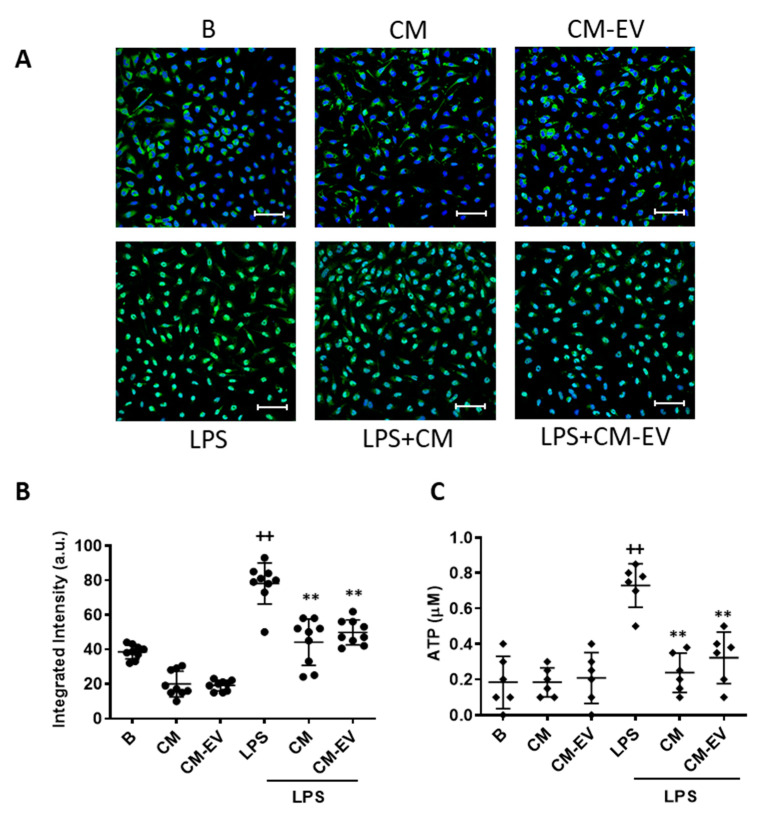
Figure 5.
NF-κB nuclear translocation and ATP release. For NF-κB translocation, macrophages were incubated with CM or CM-EV for 30 min and immunofluorescence was determined by confocal microscopy using an NF-κB p65 XP® Rabbit (Alexa Fluor® 488 Conjugate). (A) Representative images. Microscopic magnification of the objective lens 40 x. Bar = 50 µm. (B) Quantification of nuclear translocation. For ATP release (C), macrophages were incubated with CM or CM-EV in the presence or absence of LPS for 20 h. ATP levels were determined by luminescence in the medium. B: cells incubated with control medium and not stimulated with LPS. Data are presented as mean ± SD (n = 6–9) from 3 independent experiments. One-way analysis of variance followed by Tukey’s post hoc test; ++ p < 0.001 versus B; ** p < 0.01 versus LPS control.

