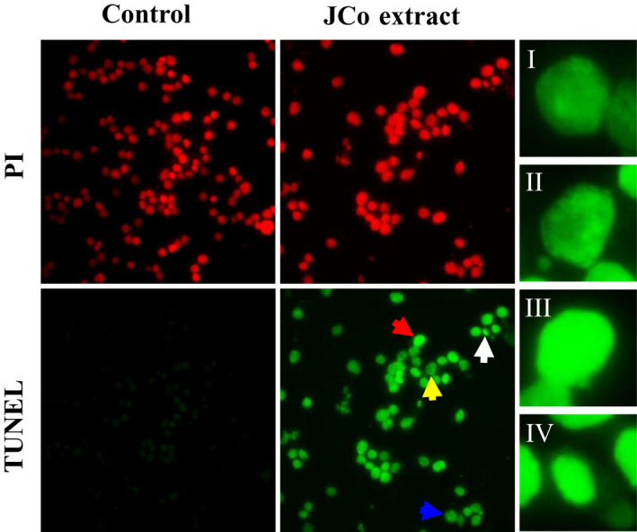
FIGURE 3.

JCo extract induced cell apoptosis in esophageal cancer cells. CE81T/VGH cells were treated with 70 μg/mL JCo extract for 48 hr, and apoptosis was determined by a TUNEL assay. In the fluorescence images, anoikis (I, blue arrow), chromatin condensation (II, yellow arrow), DNA fragmentation (III, red arrow), and an apoptotic body (IV, white arrow) are shown
