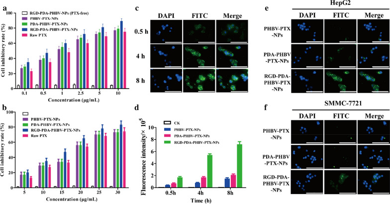
Fig. 6.
a The inhibitory effect of nanoparticles on HepG2 cells. b The inhibitory effect of nanoparticles on SMMC-7721 cells. c CLSM images of HepG2 cells incubated with RGD-PDA-PHBV-PTX-NPs for 0.5, 4 and 8 h. d The fluorescence intensity of HepG2 cells incubated with different nanoparticles for 0.5 h, 4 h and 8 h. CLSM images of HepG2 cells (e) and SMMC-7721 cells (f) incubated with different nanoparticles for 4 h. Scale bar: 50 μm. PHBV, ploy (3-hydroxybutyrate-co-3-hydroxyvalerate); PTX, paclitaxel; PDA, polydopamine; NPs, nanoparticles

