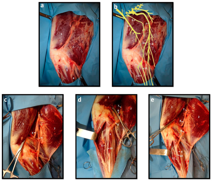
Figure 1.
Anatomical and topographic distribution of sheep’s hind limb nerves. (a) Muscles of the sheep’s hind limb—lateral view; (b) Schematic representation of the topographic distribution of the main nerves in the sheep’s hind limb; (c) Muscles and nerves of the sheep’s hind limb—deep exposure of the proximal region; (d) Muscles and nerves of the sheep’s hind limb—deep exposure of the distal region; (e) Muscles and nerves of the sheep’s hind limb—deep exposure. (1. M. vastus lateralis; 2. M. biceps femoris; 3. M. extensor digitalis lateralis; 4. M. peroneus longus; 5. Femoral n.; 6. Obturator n.; 7. Sciatic n.; 8. Tibial n.; 9. Common peroneal n.; 10. Superficial peroneal n.; 11. Deep peroneal n.).

