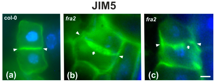
Figure 10.
DeSPHG localization by the JIM5 (green) antibody in post-cytokinetic cells of wild-type (a) and fra2 (b,c) roots. DNA counterstaining appears blue. JIM5 fluorescence reveals that the consolidated cross wall (defined by arrowheads in all figures) is evenly thick in the wild-type (a), while in fra2 it appears unevenly thick (arrow in (b)) or with gaps (arrow in (c)). Bars: 10 μm.

