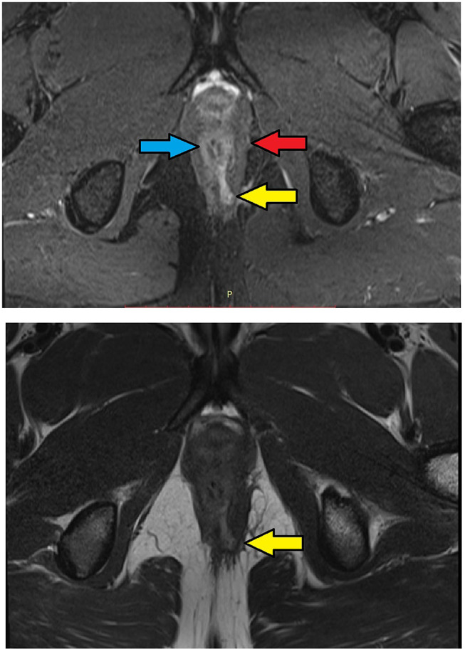
Figure 3.

MRI-axial sections in a 30-year-old male with low transsphincteric anal fistula at 5 o’clock position. The tract traverses through both external and internal anal sphincters and opens in the anal canal at the posterior midline position. Upper panel – T2, lower panel – STIR (Yellow arrows are showing fistula tract, blue arrow are showing the internal sphincter, red arrow are showing the external sphincter).
