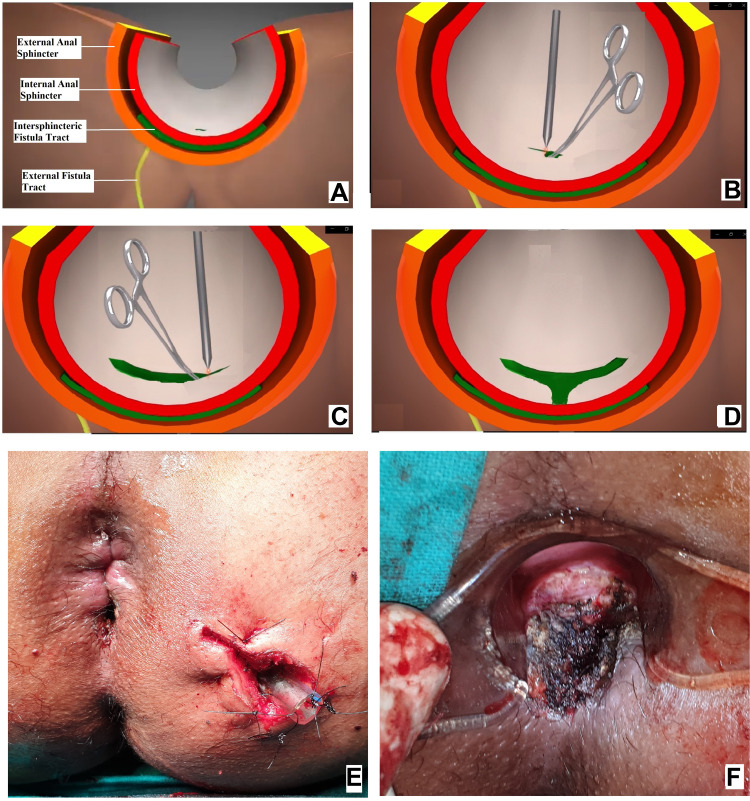
Figure 8.
Intraoperative wound after TROPIS (transanal opening of intersphincteric space). (A) Schematic diagram of anal fistula and anal canal, (B) Schematic diagram showing the intersphincteric portion of the fistula tract (green colour) and an artery forceps inside the internal opening about to be laid open with electrocautery, (C) Schematic diagram showing the intersphincteric portion of the fistula tract laid open with electrocautery, (D) Intersphincteric space distal (inferior) to the internal opening laid open by a vertical incision, (E) Intraoperative photograph of a patient after a complete TROPIS procedure showing the TROPIS wound in the anal canal and a tube inserted in the tract in left ischiorectal fossa. The tube sutured to the skin with monofilament non-absorbable suture (2–0 nylon). (F) Intraoperative photograph showing the TROPIS wound in the anal canal.

