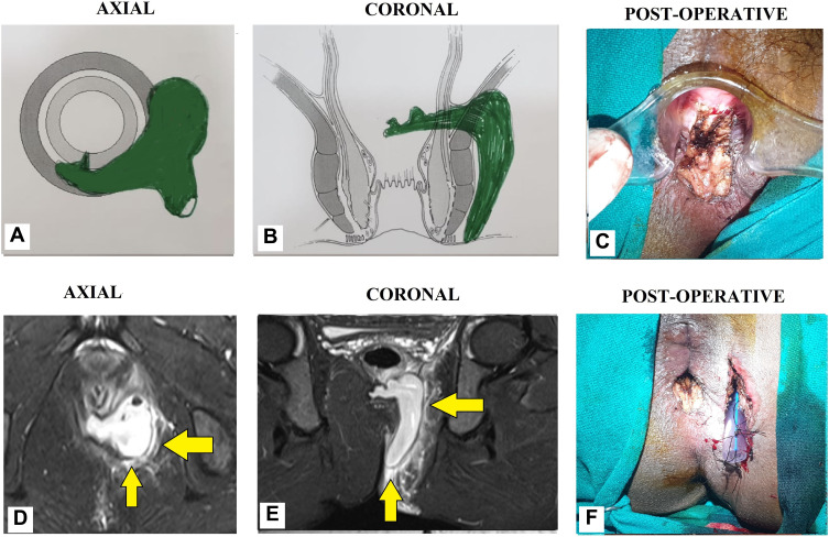
Figure 10.
A 34-year-old male patient with a high horseshoe transsphincteric anal fistula and abscess. (A): Axial section (Schematic diagram), (B) Coronal section (Schematic diagram), (C) Post-operative photograph showing TROPIS wound (laid open intersphincteric portion of the fistula tract) in the anal canal. (D) MRI-axial section (STIR), (E) MRI-coronal section (STIR), (F) Post-operative photograph showing the final picture (TROPIS wound in the anal canal) and a tube inserted in the tract in left ischiorectal fossa. The tube sutured to the skin with monofilament non-absorbable suture (2–0 nylon) (Yellow arrows are showing fistula tracts).

