Figure 5.
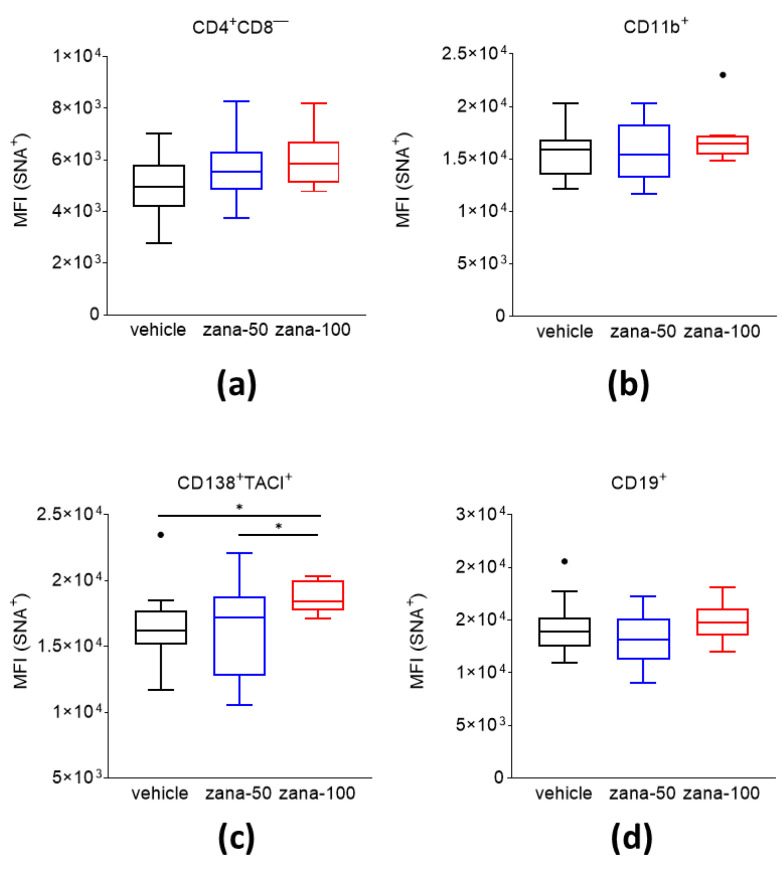
An increased level of alpha-(2,6)-sialic acids is observed on the cell surface of CD138+/TACI+ plasma cells after zanamivir treatment in CIA. Levels of (2,6)-SA residues on cell subsets—(a) CD4+CD8−T cells, (b) CD11b+ cells, (c) CD138+/TACI+ cells, and (d) CD19+cells—were measured by flow cytometric analysis. Data are presented as Tukey’s box- and whisker plots. Observations outside the whiskers are considered outliers and presented as black dots. * p < 0.05. Welch’s ANOVA test with Dunnett’s T3 multiple comparisons was used for statistical analysis.

