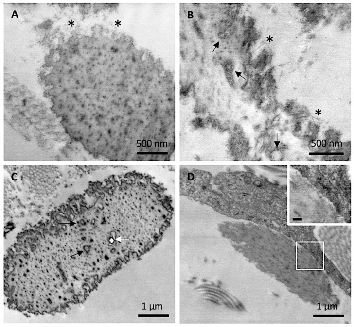
Figure 3.
Representative electron microscopy images obtained from clinically unaffected skin. (A,B) Elastic fibers at the periphery exhibit numerous microfibrillar structures (*) whereas, in the amorphous core, round electron-translucent structures are visible (black arrows). (C) Round electron-translucent structures (black arrows) as well as empty holes (white arrow) are visible. (D) A dermal fibroblast in close contact with elastic fiber. Insert bar = 100 nm.

