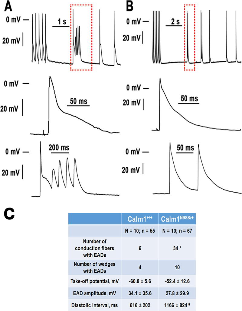
Figure 8. Pause-dependent early afterdepolarizations in the His-Purkinje network of Calm1N98S/+ hearts.
A and B Top: original traces of membrane potential obtained from His-Purkinje myocytes in Calm1N98S/+ hearts. The traces shown include the last 5 action potentials from a train of 30 action potentials at 5 Hz pacing, followed by >1-s pauses and spontaneous action potentials. Middle: Magnified views of the final paced action potential. Bottom: Magnified views of the boxed regions in the top panels, illustrating pause-dependent repetitive early afterdepolarizations (A) and an early afterdepolarization (B) arising from a spontaneous action potential. Neither cell developed diastolic (delayed) afterdepolarizations. Note the presence of automaticity in the fiber shown in B. C, Properties of early afterdepolarizations. N and n are the number of hearts and conduction fibers, respectively. EAD, early afterdepolarization; mean values for take-off potential, EAD amplitude and diastolic interval preceding the EAD-containing cycle were averaged from 202 and 23 EADs in Calm1N98S/+ and Calm1+/+ wedges, respectively. * P < 0.001 versus Calm1+/+ by Chi-square test. # P = 0.006 versus Calm1+/+ by Mann-Whitney Rank Sum test.

