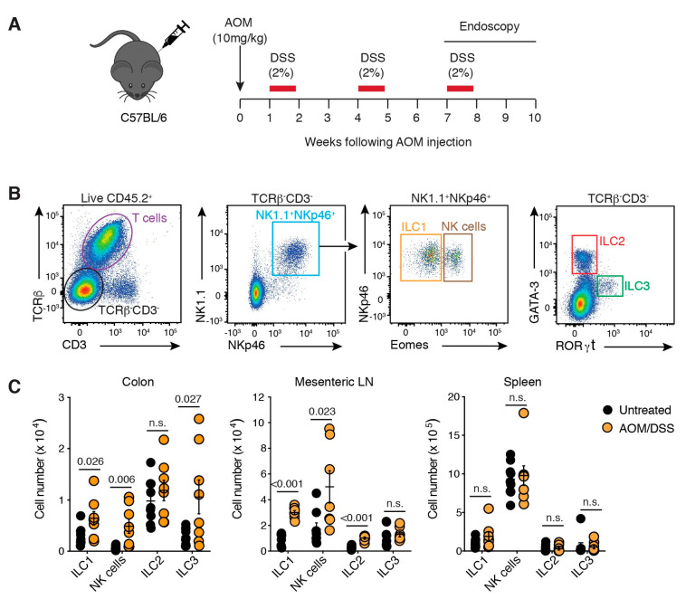
Figure 1.
ILCs infiltrate colorectal tumours. (A) Schematic illustration of the AOM/DSS treatment protocol. Naïve C57BL/6 mice injected with AOM (10 mg/kg, i.p.) followed by three five-day cycles of 2% (w/v) DSS ad libitum in their drinking water separated by two weeks of normal water between each cycle. Tumours developed in the distal colon between 7–10 weeks after the commencement of treatment. (B) Flow cytometric analysis of ILCs within lamina propria isolated from the colon. Live CD45.2+ lymphocytes were segregated into T cells (TCRβ+CD3+) and ILCs (TCRβ−CD3−). ILC subsets were further delimited into NK1.1+NKp46+ ILC1 (Eomes−) and NK cells (Eomes+); ILC2s (GATA-3+) and ILC3 (RORγt+). (C) Enumeration of ILC subsets isolated from the colon, mesenteric lymph node (LN) and spleen of untreated and AOM/DSS-treated mice 7–10 weeks after initial treatment. Each dot represents one mouse. Data show mean ± s.e.m of results pooled from four independent experiments (n = 2–3 mice/treatment/experiment). Statistics were calculated using an unpaired Student’s t-test, p-value as indicated. n.s.: not significant.

