Figure 1.
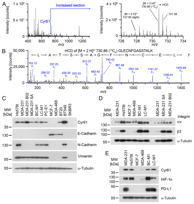
Detection of Cyr61 in tumor cell lines with mesenchymal attributes. (A) Detection of Cyr61 by SILAC LC-MS/MS. Left: MS1 (survey scan) mass spectrum containing the peptides around m/z 731 Da. Right: enlarged section of the Cyr61 peptides containing the light masses from MDA-MB-468 and the 13C6-labelled peptides of BC-M1. (B) Positive ion mode HCD (higher-energy collisional dissociation) spectrum of m/z 730.86 [M + 3 H]3+ Da. The relevant fragments of the y-ion series are assigned with their masses. (C) Confirmation of the differential expression of Cyr61 and comparison with the epithelial grade in breast cancer cell lines or DTC cell lines (BC-M1, LC-M1, PC-E1) by Western blot analysis. (D) Detection of the integrin species in the breast cancer cell lines by Western blot analysis. (E) Comparison of the protein levels of Cyr61, HIF-1α and PD-L1 in the breast cancer cell lines, as detected by Western blot analysis. (A,B) nbiol = 4; (C–E): nbiol = 3.

