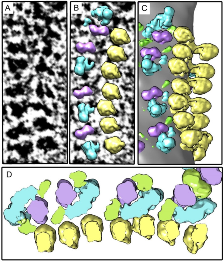
Figure 2.

An example of the oligomeric linear structure consisting of tightly docked ATP synthases and respirasomes. Elongated transmembrane parts of complexes I are oriented approximately parallel to the row of ATP synthases. (A) Tomographic slice; (B) Density maps of complexes I, III2, and ATP synthases placed back to the tomogram; (C) Surface rendering of the selected area; (D) Slice of the density map, view from the intermembrane space. Image (D) is a bit enlarged in comparison with images (A–C). Colors: yellow–ATP synthase, blue–complex I, purple–complex III dimer, green–complex IV.
