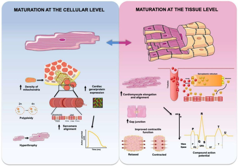
Figure 2.
General features of cardiac maturation at the cellular and tissue levels. Cardiac maturation at the level of cardiomyocytes (blue-shaded background) can be experimentally ascertained by a more adult-like cardiac gene expression profile, changes in sarcomeric ultrastructure resulting in sarcomere elongation and alignment, increased mitochondrial density to accommodate for higher energy demand, and massive growth in cardiomyocyte size (hypertrophy) with increased percentage of polyploid cells. Electrophysiologically, cardiomyocyte maturation results in a more negative resting membrane potential and increased duration and amplitude of action potential. Cardiac maturation at the tissue level (pink-shaded background) is characterized by elongated cardiomyocytes preferentially aligned along a specific axis, increased expression of Cx43 and distinct localization of gap junction proteins, and improvements in impulse propagation and synchronization of cardiac contraction, leading to cardiomyocytes performing as a functional syncytium. Maturation at the tissue level results in increased contractility and force generation and, electrophysiologically can be registered as compound action potentials reminiscent of surface ECG recordings. Figure created with BioRender.com and smart.servier.com.

