Figure 1.
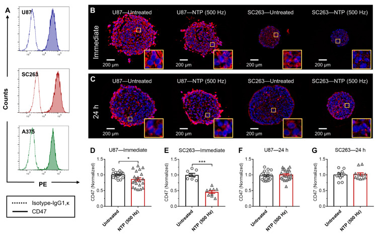
NTP treatment modulated CD47 immediately in 3D tumor spheroids. (A) The baseline expression of three human cancer cells, glioblastoma (U87), head and neck squamous cell carcinoma (SC263), and melanoma (A375) was analyzed using immunohistochemistry and flow cytometry. U87 and SC263 cells were able to form compact spheroids and were exposed to NTP. Spheroids were collected (B) immediately or (C) 24 h after NTP treatment, paraffin-fixed, sectioned, stained for CD47 (red), and counter-stained with a nuclear dye, 4′,6-diamidino-2-phenylindole (DAPI) (blue). Images were taken together at fixed microscope settings (10×) per experiment and all images were batch processed. Yellow inserts are a zoomed-in area (100 µm × 100 µm) to show CD47 staining surrounding the nucleus. CD47 expression was quantified and normalized to the untreated (D–G). Data here are represented as mean ± SEM and individual values are also shown (n = 8–21). Statistical significance of all treatment conditions was determined using the generalized linear mixed model with post hoc Dunnett’s test comparison to untreated (* p ≤ 0.05; *** p ≤ 0.001).

