Figure 1.

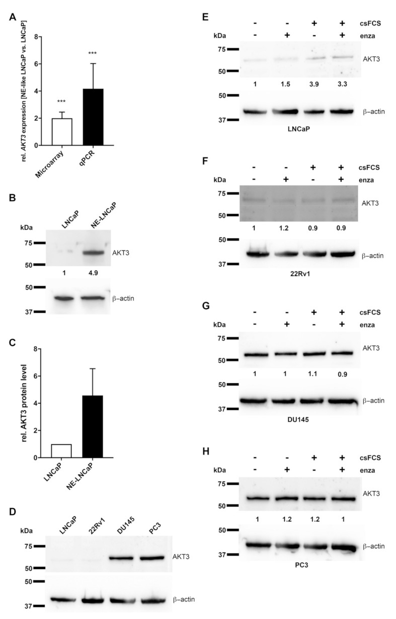
Expression of AKT3 is increased in neuroendocrine (NE)-like LNCaP cells and inhibited by androgen receptor (AR) signaling in prostate carcinoma (PCa) cells. (A) According to the results of microarray data from Dankert et al. 2018 (white bar; p = 0.0002) AKT3 expression was validated by qRT-PCR (black bar; p = 0.0007) confirming increased AKT3 expression in NE-LNCaP compared to normal LNCaP cells on mRNA level (*** p < 0.001). (B) Representative western blot analysis using anti-AKT3 mAb confirms induced AKT3 protein level in NE-like LNCaP cells. (C) Densitometrical quantification of western blot analyses represents the relative upregulation (4.5-fold) of AKT3 protein in NE-like LNCaP cells, determined in five independent experiments in relation to corresponding β-actin as loading control. (D) Western blot analysis of four PCa cell lines using anti-AKT3 mAb depicts different AKT3 expression in AR-positive and AR-negative PCa cell lines. PCa cell lines were treated with androgen deprivation or 10 µM enzalutamide followed by western blot assays for AKT3 expression. The treatments increased AKT3 protein expression in LNCaP cells (E), whereas AKT3 expression did not change in 22Rv1 (F), DU145 (G) and PC3 (H) cells.
