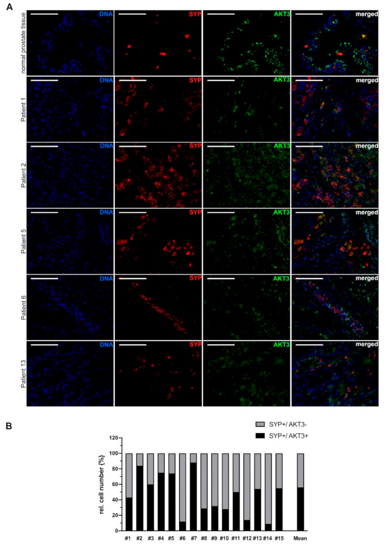
Figure 3.

AKT3 colocalization with NE cell marker SYP in advanced prostate cancer tissue. (A) Immunofluorescence double staining of normal prostate tissue and representative tissue samples from 5 patients with neuroendocrine differentiated prostate carcinoma showing SYP-positive NE cells expressing AKT3. AKT3 frequently is colocalized with SYP in normal prostate tissue as well as tissue samples from patients with neuroendocrine PCa, indicating expression of AKT3 in NE cells (DNA: blue, CHGA-positive NE-cells: red, AKT3: green, double positive cells: yellow; Magnification: 400×, scale bar: 100 µm). (B) Three areas per patient with neuroendocrine-differentiated prostate cancer were documented and only samples with more than 20 neuroendocrine cells were analyzed. The number of SYP-positive NE cells was normalized to 100% (grey bar). Graph shows statistical evaluation that demonstrates colocalization of SYP and AKT3 (black bar) in approximately 55% of neuroendocrine cells.
