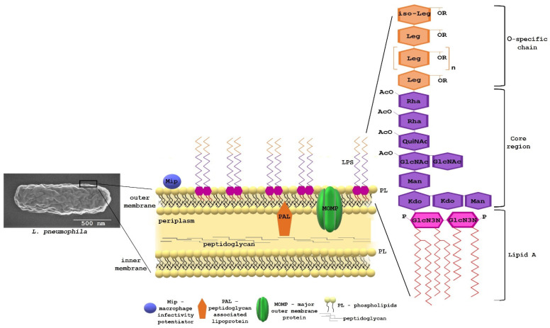
Figure 1.
Scanning electron microscopy image of Legionella pneumophila, and model of the L. pneumophila cell envelope. Structure of lipopolysaccharide (LPS) (modified from [16]). Leg., legionaminic acid; Rha, rhamnose; OAc, O-acetyl; QuiNAc, acetylquinovosamine; GlcNAc, acetylglucosamine; Man, mannose; Kdo, 3-deoxy-d-manno-oct-2-ulosonic acid; P, phosphate; and GlcN3N, 2,3 diamino-2,3-dideoxy-d-glucose.

