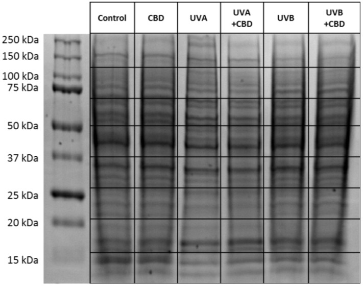
Figure 9.
SDS–PAGE separation and staining with Coomassie brilliant blue R-250 of proteins from control keratinocytes and irradiated with UVA (30 J/cm2), UVB (60 mJ/cm2) or/and treated with cannabidiol (CBD, 4 μM) in a three-dimensional (3D) culture model. The grid indicates the borders of the protein migration zones. Original photo of the gel is added in Supplementary Figure S1.

