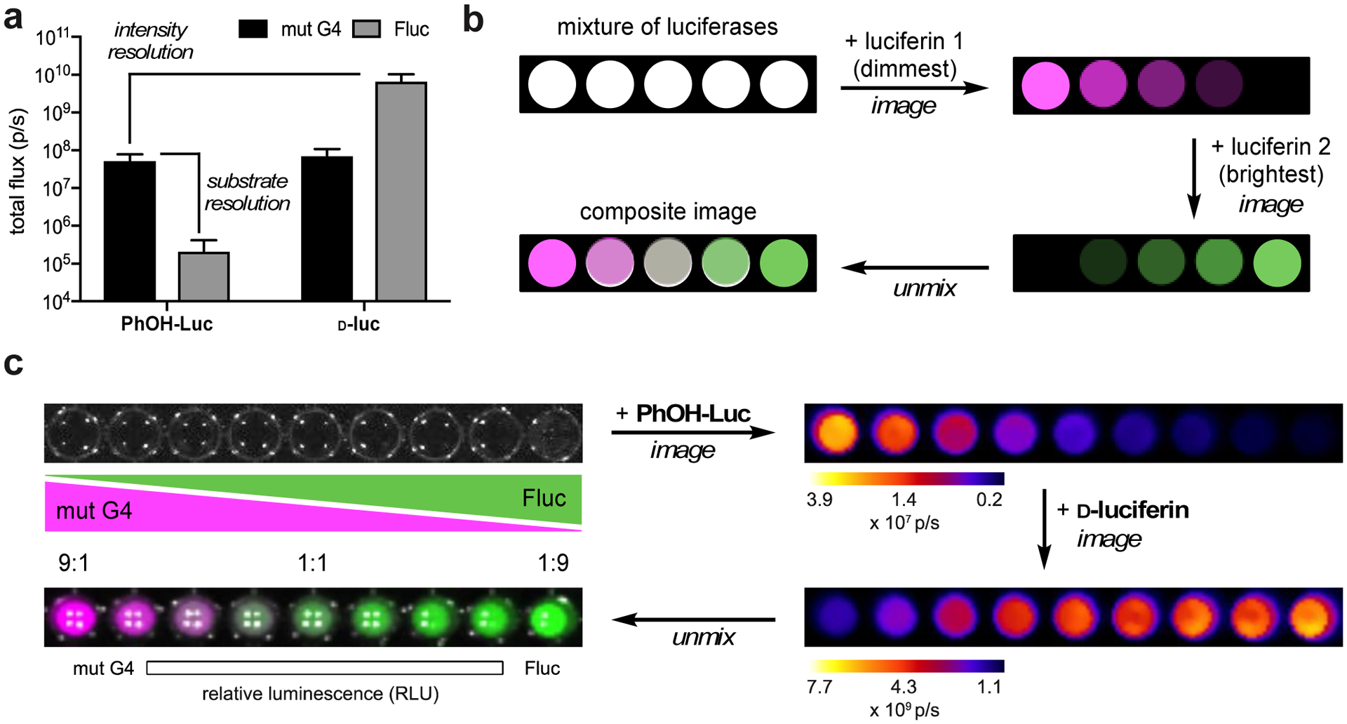
Figure 5. Rapid, two-component imaging with a π-extended luciferin.

(a) G4/PhOH-Luc are both substrate- and intensity-resolved from Fluc/d-luc. (b) Schematic for resolving multiple luciferases via substrate unmixing. Luciferin analogs were administered sequentially with minimal delay time. Bioluminescent signals generated from each addition were deconvoluted via linear unmixing. (c) A model two-component assay. Gradients of G4 and Fluc (expressed in bacterial lysate) were plated in a 96-well plate. PhOH-Luc (250 μM) was administered, followed by d-luc (100 μM). Images were acquired after each addition. The raw data were stacked and unmixed. The processed image is false colored.
